Human Eye Mode
Product Details:
- Automation Grade Manual
- Power Source None
- Warranty 6 months manufacturing defect warranty
- Temperature Range Room temperature
- Model No EDEX-HEM123
- Feature Detailed anatomical representation
- Core Components Eye model parts
- Click to View more
X
Human Eye Mode Product Specifications
- EDEX-HEM123
- Manual
- Room temperature
- Physical model
- Standard dimensions
- Standard lightweight
- 6 months manufacturing defect warranty
- Plastic Painted Parts
- None
- Anatomical Model
- Eye model parts
- Detailed anatomical representation
- Educational purposes medical training
Product Description
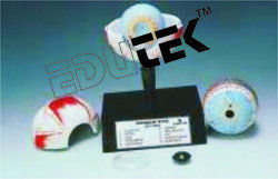
Human Eye Model
HUMAN EYE, 3 TIMES ENLARGED
An highly improved and enlarged model showing fine details in three dimensional relief. Cut in horizontal plane and separable in seven parts. After removing the upper half of the sclera (outer shell), the choroid with its verticose veins is exposed; with the removal of second shell, details of the retina come into view and the position of the yellow and blind spots is quite visible. All important anatomical features such as muscle insertions, optic nerve, blood vessels, ciliary body, cornea, crystalline lens, and iris etc are clearly numbered and are identifiable. Mounted on base, with key card.
FAQs of Human Eye Mode:
Q: What is the temperature range suitable for the Human Eye Model?
A: The Human Eye Model is designed to be used at room temperature.Q: What are the core components of the Human Eye Model?
A: The core components of the Human Eye Model include eye model parts.Q: What type of display does the Human Eye Model have?
A: The Human Eye Model features a physical model display.Q: What is the warranty period for the Human Eye Model?
A: The Human Eye Model comes with a 6 months manufacturing defect warranty.Q: What is the power source required for the Human Eye Model?
A: The Human Eye Model does not require any power source.Q: What is the primary usage of the Human Eye Model?
A: The Human Eye Model is primarily used for educational purposes and medical training.Q: What is the material used for the Human Eye Model?
A: The Human Eye Model is made of plastic painted parts.Tell us about your requirement

Price:
Quantity
Select Unit
- 50
- 100
- 200
- 250
- 500
- 1000+
Additional detail
+91
Email
Other Products in 'Biology Equipments' category
"We deal all over World but our main domestic market is South India"
 |
EDUTEK INSTRUMENTATION
All Rights Reserved.(Terms of Use) Developed and Managed by Infocom Network Private Limited. |


